
Case Sharing
2022-10-25 00:00

Specialist Introduction:
Head of Urolithiasis Subspecialty Group, Xinhua Hospital affiliated to Shanghai Jiaotong University School of Medicine
Member of the Calculus Committee of the Urology Branch of the Shanghai Medical Association
Member of the Calculus Group of the Male Urology Special Committee of the Shanghai Association of Chinese Integrative Medicine
Member of Society for the Evaluation and Management of Clinical Anti-Infective Drugs, Chinese Medicine Education Association
Case Introduction:Ureterorenoscopic Lithostripsy (stage I) via the left ureter
Basic information
• Female, 63 years old
• Main Complaint: Left lower back pain with vomiting for more than 3 months
• Past Medical History: PCI was performed for coronary artery disease in 2014 and again in 2016 for a recurrence of coronary artery disease. She denied a history of other surgical procedures, trauma, and blood transfusion.
• History of Present Illness: Approximately three months ago, the patient developed pain and discomfort in the left lower back without an obvious cause, which was paroxysmal and radiated to the lower abdomen, accompanied by vomiting, without fever, chills, nausea, and acid reflux, and denied hematuresis, frequent urination, urinary urgency, feeling of endless urination, and pain in urination. The patient was then treated in another hospital, where a CT examination showed left-sided renal calculi (the specific report is not available). The symptoms were relieved with medication and then the patient experienced repeated left low back pain and discomfort. Approximately 20 days ago, the patient again presented with left lumbar pain, which was worse than before and persisted without relief, and she came to our emergency clinic. Urine routine of our hospital (2022/08/20): erythrocyte hemoglobin 150/uL↑, erythrocyte (microscopy) full field/HP; Abdominal CT of our hospital (2022/08/20): bilateral renal calculi, especially with left kidney, left renal hydronephrosis, possible cyst in the right kidney, please refer to the clinical situation. Small hypodense foci in the spleen, follow up is recommended. The patient was admitted for further treatment of the stones and is accepted with "Bilateral Renal Calculi". Since the onset of the disease, the patient has been conscious and mentally competent, with an average appetite and nighttime sleep, no difference in bowel movements, and no significant weight loss.
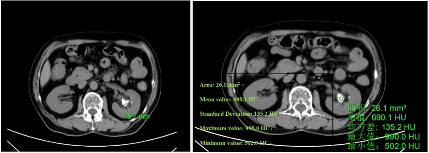
• CT:Partially cast stones in the left kidney 1.5×3.5cm
Treatment process
• Surgery Name: Ureterorenoscopic Lithostripsy (stage I) via the left ureter
• Surgical Procedure:
1. Lithotripsy clearance is performed using Scivita Medical's Single-use Urology Videoscope in combination with curvable negative pressure sheath from YigaoMed.
2. Place a 200 μm holmium laser fiber with an energy setting of 1.0 J at 20 Hz to powder and fragment the stones, with a negative pressure sheath against the fragmented stone to adsorb them out of the body.
3. After the stone has been completely removed, place a guide wire in the sheath, and use Scivita Medical's Single-use Urology Videoscope to fully examine the renal collecting system and observe the renal pelvis and calyces for residual stones.
Surgery Summary:
1. The patient had double kidney stones with heavy stone load, and might need longer time to remove the stone after lithotripsy. With residual stones, there is the possibility of needing a second operation to remove the stones.
2.The heavier the stone load of the patient, the higher the ratio of powdered fragmental stones during operation, the higher of the probability of clearing away all the stones, the shorter the time to remove stones after operation.
3. Larger stones should be moved to the upper calyces during operation, and then be smashed, thus it helps the removal of residual stones after operation.
4.For the stones with heavier load, lower calyces stones, difficult stones (such as the stones in diverticulum), disposable soft lithotriptoscepe can be first considered, so as to reduce the damage on the high value reusable electronic lithotriptoscepe, as it has high definition and operability and can also improve the effect of the operation.
5. Definite diagnosis on the operation on difficult stones ( such as special anatomical structure, special patient condition, special stone location, etc ) before operation, and sufficient preparation, can lower intraoperative risk and the possibility of failed operation, and improve operation effect and safety.

Contact phone number:
Company address:
No. 2, Qingqiu Street, Suzhou Industrial Park, Suzhou, Jiangsu Prov., P.R.CHINA,215127

Copyright © Scivita Medical All Rights Reserved
网站备案/许可证号:苏ICP备18001379号-1
互联网药品信息资格服务资格证书编号:(苏)-非经营性-2022-0018